Everything ECMO 001
Author: Chris Nickson
Reviewers: Vin Pellegrino and Jayne Sheldrake
A 26 year-old man with severe ARDS due to influenza is currently receiving VV ECMO for respiratory failure.
You are asked to review him because an arterial blood gas (ABG) from his right radial arterial line shows that he has an Sa02 of 80%, which is consistent with the SpO2 monitor on his left index finger.
Q1. What 4 things should you check initially?
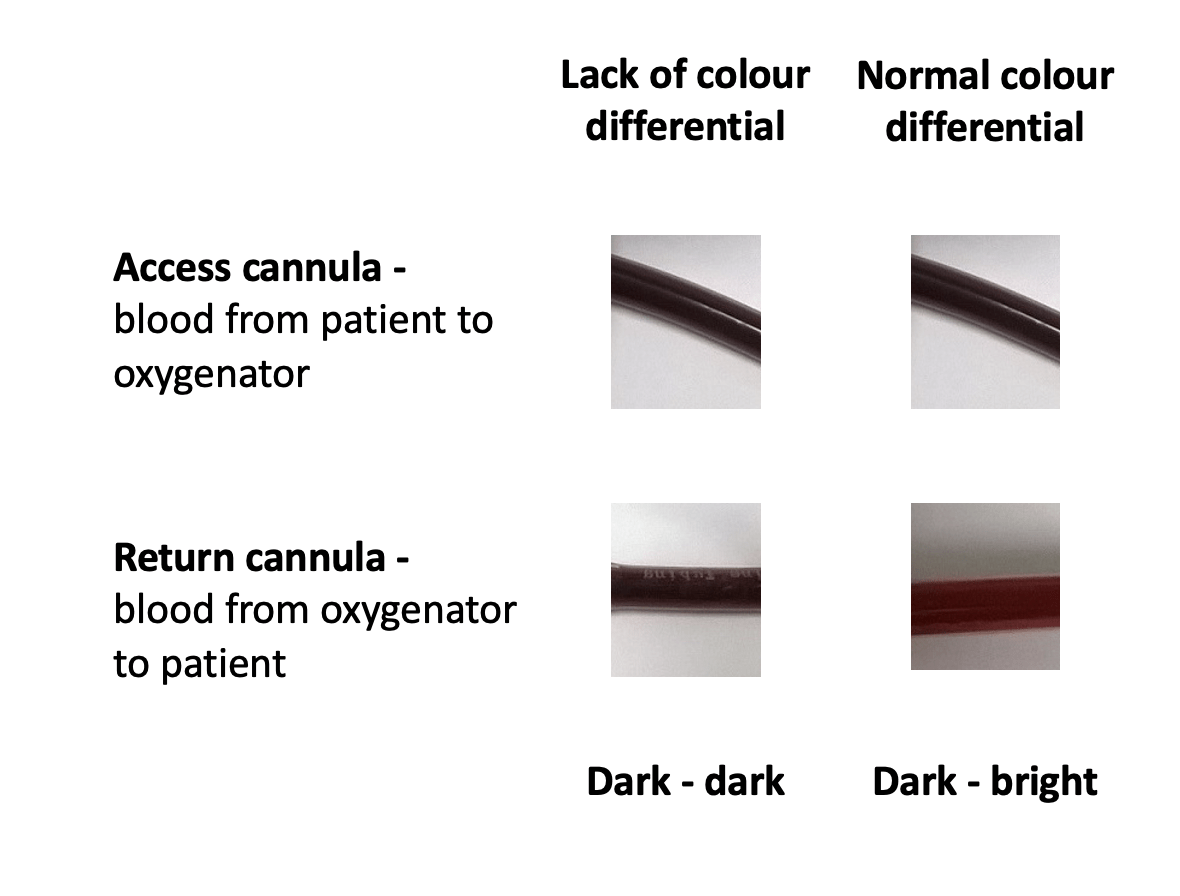
- presence of colour differential between access and return cannula
- fresh gas flow (FGF)
- Blender
- Flow calibrations
The first checks to make in a patient on VV ECMO are to ensure adequate oxygen delivery to the oxygenator and appropriate circuit blood flow.
On initiating VV ECMO the FGF and the ECMO blood flow are usually commenced at a 1:1 ratio and then adequacy is checked by performing an ABG.
Before anything else, quickly check for colour differential between the access and return cannula. If the blood being returned to the patient from the oxygenator is as dark as the blood entering the oxygenator, then oxygen supply failure or oxygenator failure are likely.
Ensure that the fresh gas tubing is connected to the inlet from the fresh gas flowmeter. FGF is titrated to the target PaCO2. However, if the patient is hypoxaemic it is important to check that the FGF has not been excessively decreased (read from the center of the ‘ball’ on the flowmeter). Don’t confuse the high and low flow sides of the fresh gas flowmeter (make sure only one is selected).
At The Alfred ICU the FGF blender setting is routinely set at 100% O2 and is not usually changed, even during weaning. This guards against unintentional inadequate oxygen delivery to the oxygenator.
Thirdly, flow calibrations should also be checked. This can be done by turning the pump speed (rpm) to zero while the circuit is clamped, then holding down the ‘0’ button on the PLS or pressing the zero indicator button on the HLS. Remember to then increase pump speed to 1000 rpm before slowly releasing the clamp over 3-5 seconds and then continuing to slowly increase the rpm until target flow is achieved.
Target blood flows are chosen such that adequate arterial oxygenation is achieved while allowing non-injurious lung ventilation.
Your checks found nothing untoward. The low SaO2 persists.
Q2. What should you check next?
The next step is to check for access insufficiency.
Suspect access insufficiency if any of the following are present:
- unstable circuit flows
- rising negative pressures (if using the HLS system)
- kicking or swinging of the venous drainage line
Access insufficiency can be confirmed by performing a ‘ramp test’: increase the pump speed (rpm) in a stepwise fashion and record the ECMO blood flows that are achieved:
- there is no access insufficiency if ECMO blood flow steadily increases with increasing rpm
- there is access insufficiency if ECMO blood flow decreases once a threshold rpm is exceeded
Access insufficiency, if present, must be corrected before you do anything else (a topic for another post!).
Despite the further checks, no cause is found. The patient remains hypoxaemic.
Q3. What will you do next, and why?
Having excluded access insufficiency, you can now increase the circuit blood flow (typically to 6-7 L/min in adults). Normally functioning oxygenators are efficient at oxygenating blood so this should correct any V/Q mismatch caused by insufficient circuit flows.
You do that… The patient is still hypoxaemic!
Q4. Now what will you do, and why?
Perform a pre-oxygenator blood gas to check the SO2.
Having excluded many of the possible causes of hypoxaemia in a VV ECMO patient, we still need to consider the possibility of high oxygen consumption or recirculation.
- Low SO2 (<60%) suggests high oxygen consumption
- High SO2 (>80%) suggests recirculation
Q5. What are your options if the measurement performed in Q4 is LOW?
If there is high oxygen consumption your options include:
- Tolerating the decreased SaO2
- Target a higher haemoglobin to increase oxygen delivery (e.g. Hb >100 g/L)
- Improve oxygenation through the lungs (e.g. try nitric oxide or prostacyclin)
- Commence neuromuscular blockade to decrease oxygen demand
- Use targeted temperature management to cool the patient and decrease oxygen demand
Q6. What are your options if the measurement performed in Q4 is HIGH?
This suggests that recirculation is occurring. If so the access cannula SO2 will be high, despite the low SO2 from the patient’s arterial line. It means that flow from the return cannula is entering the access cannula, rather than being distributed to the patient.
Check cannula positions
- see if there has been displacement of the cannulae at the insertion site
- check position of tips on x-ray
Regardless of the configuration of VV ECMO (femoral-femoral, femoral-jugular or high flow) the tip of the access cannula should be 8 to 15cm apart to minimise recirculation.
An alternative explanation is that the cannula are correctly positioned but flowing in reverse… Make sure this isn’t the case!
Reference
- Pellegrino V, Sheldrake J, Murphy D, Hockings L, Roberts L. Extracorporeal Membrane Oxygenation (ECMO). Alfred ICU Guideline, 2012.


Pingback: Everything ECMO! | Edwin M. Thames