Labs and Lytes 025
Author: Chris Sia
Reviewers: Sarah Yong and Chris Nickson
A 30 year-old lady presents with acute type 1 respiratory failure and fever
Click image to enlarge
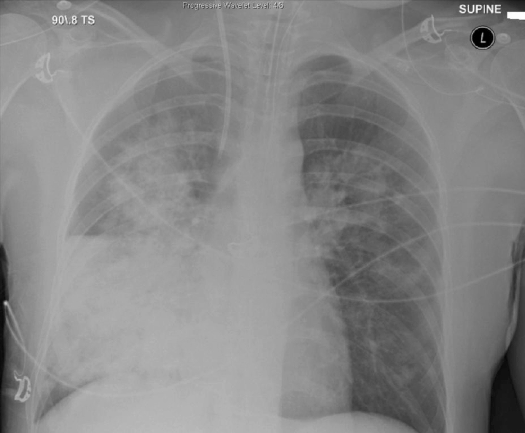
Q1. Describe the x-ray findings?
This is an AP supine CXR.
The most striking findings are bilateral alveolar opacities in the upper zones and a confluent opacity in the right middle and lower zones.
Other features:
- Right internal jugular central line
- Intubated
- Nasogastric tube (difficult to determine if adequately positioned on this image)
- Obscuration of the right heart border and preservation of the diaphragm suggests RML involvement
Q2. What are the causes of alveolar (airspace) opacities?
Causes include:
- Fluid: cardiogenic and non-cardiogenic pulmonary oedema, ARDS, aspiration
- Pus: Pneumonia (bacterial, atpical, fungal, viral, parasitic)
- Blood: Trauma (contusion), immunological (Goodpasture’s syndome), bleeding diathesis (coagulopathy), pulmonary embolism
- Protein: Alveolar proteinosis
- Cells: Adenocarcinoma in situ (bronchoalveolar cell cancer), lymphoma
Alveolar or airspace abnormalities implies different undlerlying causes to interstitial opacities.
Q3. What are the differentials of interstitial opacities?
Upper zone: SCART
- Silicosis/Sarcoidosis
- Coal workers pneumoconiosis
- Ankylosing spondylitis, allergic bronchopulmonary aspergillosis
- Radiotherapy
- Tuberculosis
Lower zone: RASIO
- Rheumatoid arthritis and other connective tissue diseases
- Asbestosis
- Scleroderma
- Idiopathic pulmonary fibrosis
- Other: Drugs (eg methotrexate, amiodarone, bleomycin, hydralazine, amiodarone, nitrofurantoin, busulphan)
In general, inhalational pathologies involve the upper zone, whereas many systemic (‘blood-borne’) pathologies involve the lower zones
References and links
- LITFL: Pulmonary fibrosis DDx
- LITFL: Pulmonary opacities on CXR
Awesome information